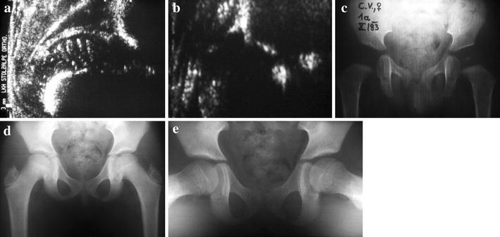
Fig. 4.

Typical clinical follow-up after early sonographic diagnosis. a Diagnosis of a highly decentered sonographic type 4 hip at the age of 3 weeks; treatment by closed reduction and squatting cast. b Well (re-)centered sonographic type 2b hip at the age of 3 months, still under functional biomechanical treatment using a removable brace in squatting position. c X-ray at the age of 1 year: sufficient bony maturation and free of AVN. d, e X-ray at the age of 9 years in anterior-posterior (d) and axial (e) views: symmetric bony development in both projections is shown
