Fig.2.

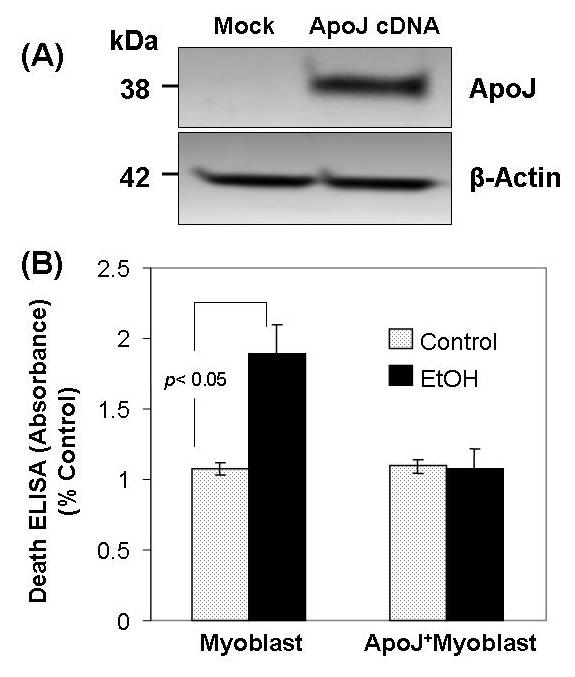
Apo J expression transduced by cDNA transfection prevents EtOH - induced apoptosis in fetal myoblast cells. Fig. 2 A. Immunoblot analysis showing ApoJ expression in fetal myoblasts transfected with or without ApoJ. Total cellular proteins extracted from fetal myoblasts and from fetal myoblasts transfected with ApoJ were subjected to 10% SDS-PAGE, then transferred to polyvinylidene fluoride membranes, blotted with anti-Apo J and anti-β-actin antibody, and detected by enhanced chemiluminescence. Anti-β-actin antibody served as a control for equal loading. Fig. 2 B. Apoptosis of ApoJ – transfected or mock control canine myoblasts exposed to EtOH. Both fetal myoblasts transfected with or without ApoJ were exposed to 100 mM EtOH for 48 h, and apoptosis was analyzed for histone-associated DNA fragments.
