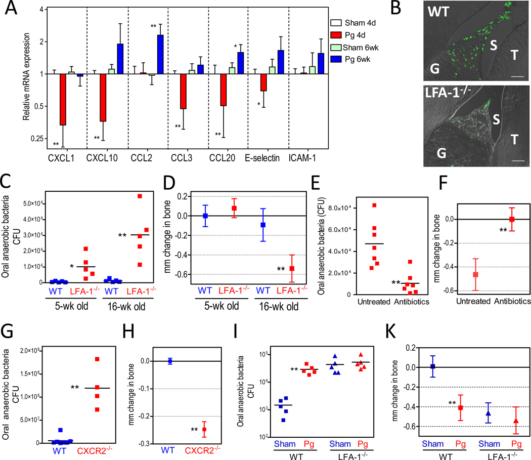
Figure 5. Compromised homeostasis in the periodontal tissue.
(A) BALB/c mice were orally inoculated with P. gingivalis (Pg) or vehicle only (sham) and sacrificed 4 days or 6 weeks post-inoculation. Dissected gingiva were processed for real-time PCR to determine mRNA expression of the indicated molecules (normalized against GAPDH mRNA and shown as fold change relative to 4-day sham-infected mice). Data are means ± SD (n = 5 mice per group). (B) Diminished recruitment of neutrophils to the gingiva of LFA-1−/− mice. Sagittal sections of periodontal tissue from 5-week-old wild-type (WT) or LFA-1−/− mice were stained for the neutrophil marker LyG6. Shown are representative overlays of differential interference contrast and fluorescent confocal images. G, gingiva; S, sulcus; T, teeth. (C–D) WT and LFA-1−/− mice were assessed for levels of oral anaerobic bacteria (C) and periodontal bone loss (D) as they aged from 5 to 16 weeks. (E–F) LFA-1−/− mice given antibiotics-containing drinking water or plain water (control) were assessed for levels of oral anaerobic bacteria (E) and bone loss (F). (G–H) Eighteen- to twenty-week-old WT and CXCR2−/− mice were assessed for oral anaerobic bacteria (G) and periodontal bone levels (H). (I–K) Ten-week-old WT or LFA-1−/− mice were orally inoculated with P. gingivalis (Pg) or vehicle only (Sham) and six weeks later were assessed for levels of oral anaerobic bacteria (I) and bone loss (K). CFU are shown for each mouse and horizontal lines denote mean values. Bone loss data are means ± SD (n = 5 to 7 mice per group). *p <0.05; **p <0.01 versus corresponding control.

