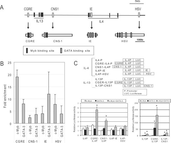
Figure 2. c-Myb regulates IL-13 and IL-4 expression through regulatory elements in the Th2 cytokine gene in cooperation with GATA-3.
(A) Schematic representation of the IL-13 and IL-4 gene locus. Exons (Black rectangles) and regulatory elements (blank rectangles) of IL-13 and IL-4 are depicted in the locus. The schemas below each arrow show the Myb (Black rectangles) and GATA binding sites (blank rectangles) in the regulatory elements. CGRE: Conserved GATA-3 Response Element, CNS-1: Conserved Noncoding Sequence 1, IE: Intronic Enhancer, HSV: Hypersensitive Site V. (B) ChIP assays were performed with anti-c-Myb and anti-GATA-3 antibodies using primary human CD4+ T cells after stimulation with IL-4, IL-2 and antibodies against CD3/CD28. Fold enrichments were measured by QRT-PCR with specific primers for CGRE, CNS-1, IE and HSV regions. All results shown (all grey bars) are statistically significant (P < 0.05) compared to control IgG. (C) Upper schema shows reporter constructs in the pGL3 vector for promoter analysis. The order of promoter and regulatory elements in the constructs is the same as the arrangement in the human gene as shown in the upper schema of Figure 1A. Graphs show the results of reporter assays for IL-13 (right) and IL-4 (left). Reporter assays were carried out in 293T cells 24 hours after transfection with each construct in the presence of c-myb and/or GATA-3 expression constructs, or control empty vectors. * Statistically significant (P < 0.05) compared to the activity of the promoter alone.

