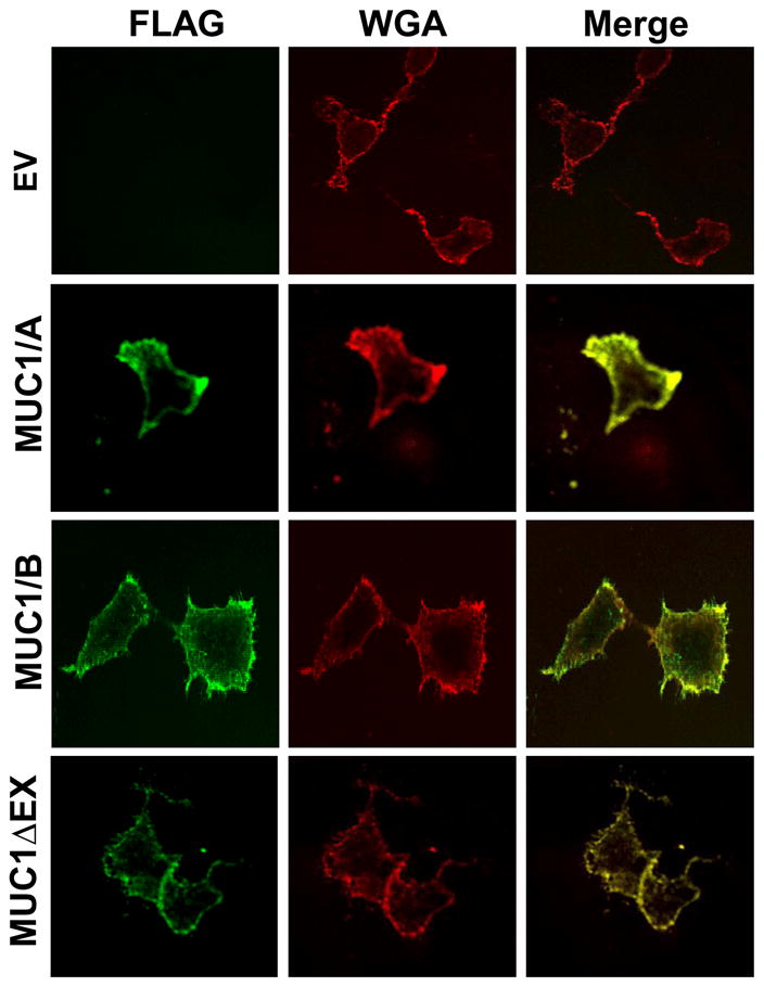
Figure 2. Surface expression of MUC1/A and MUC1/B in COS-7 cells.

COS-7 cells were transfected with the empty vector (EV, negative control), MUC1/A, MUC1/B, or MUC1ΔEX as described in Experimental Procedures. Forty-eight hours after transfection, the cells were stained with the M2 anti-FLAG antibody (green) and Wheat Germ Agglutinin (WGA, red) as a plasma membrane marker. Images were captured using an Olympus FV1000 confocal microscope. The colocalization of FLAG and WGA is indicated in yellow in the merged image.
