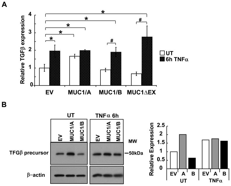
Figure 4. MUC1/A stimulates TGFβ expression.
COS-7 cells were transfected with EV control, MUC1/A, MUC1/B, or MUC1ΔEX for 48 h prior to treatment with 10 ng/ml TNFα for 6 h, UT= untreated. (A) QRT-PCR analysis of TGFβ mRNA was performed. TGFβ expression in EV-transfected/UT cells was set to one for comparison. Values are the mean ± SEM of 3 experiments. *, p<0.05 compared to EV-UT. #, p<0.05 compared to MUC1/B or MUC1ΔEX UT. (B) Western blot analysis of TGFβ precursor expression in WCE (6 μg). The blot was probed with TGFβ antibody and then stripped and reprobed for β-actin as a loading control. This blot is representative of three blots that showed the same results. The bar graph shows the quantitation of TGFβ relative to β-actin expression relative to EV that was set to one for comparison.

