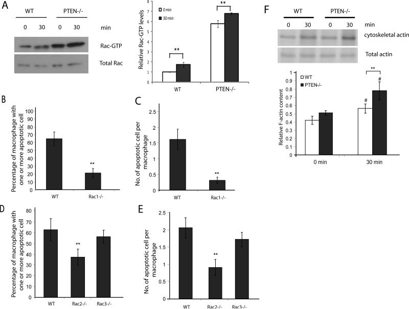
Figure 6. Increased Rac activation and F-actin polymerization in PTEN-/- macrophages during efferocytosis.
(A) Wild type or PTEN-/- peritoneal macrophages were incubated with or without apoptotic cells for 30 minutes. Unadhered cells were washed and cell lysates were analyzed for levels of activated Rac-GTPase (Rac-GTP) by GST-pulldown using GST-PAK-PBD coated glutathione beads. Cell lysates and pulldown eluates were probed for Rac1. Relative level of active GTP-bound Rac1 to total Rac1 was quantified using ImageJ and depicted below. Wild type, Rac1-/- (B-C), Rac2-/-or Rac3-/- (D-E) peritoneal macrophages were incubated with apoptotic cells at a density of (1:10) for 90 minutes at 37°C. Cells were washed and efferocytosis was analyzed by HEMA3 staining. Number of macrophages containing one or more apoptotic cell was scored as % efferocytosis, n>600 (B and D). Number of apoptotic cells engulfed by one macrophage was counted and represented as efferocytic index, n>600 (C and E). (F) Wildtype and PTEN-/- macrophages were incubated with or without apoptotic cells for 30 minutes. Unadhered cells were washed and cells were lysed in CB. Cell lysates were centrifuged and the pellets were collected, washed, and analyzed by SDS-PAGE. Graph (below) indicates mean of four independent experiments. Results shown are means ± SD. #p<0.05; **p<0.005.

