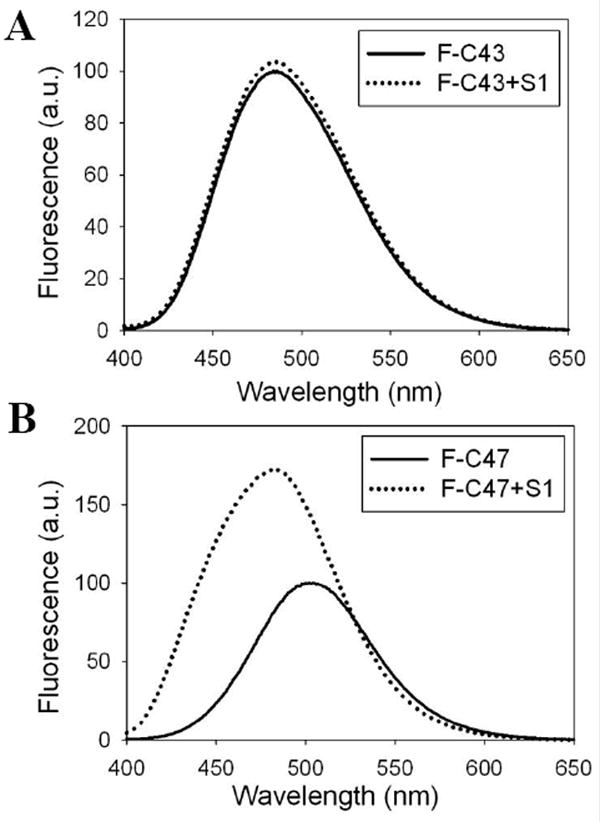
Figure 4.

Site-directed fluorescence labeling reveals myosin induced conformational changes in dynamic actin loops. Representative acrylodan emission spectra of yeast F-actin mutants labeled at residue 43 and 47 in the absence (solid lines) and presence (dotted lines) of S1 are given in 4A and 4B, respectively.
