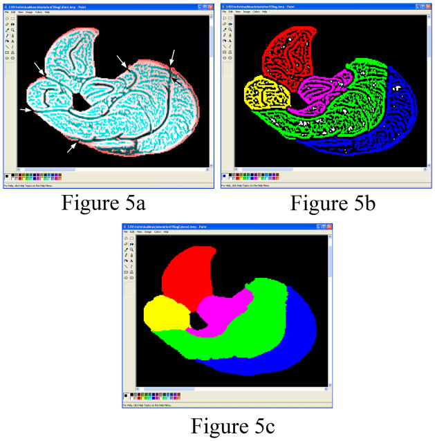
Figure 5.
(a) Edges of the individual muscles are automatically identified by the program. The user is required to break (arrows) the few small connections between the individual muscles using the Paint program’s black pencil tool. (b) Each of the individual muscles is filled using the Paint program’s ”Fill With Color” tool when the user selects a muscle. The anterior compartment is colored red, lateral yellow, deep pink, soleus green and gastroc blue. (c) After using the ”Fill With Color” tool, the muscle mask for each compartment is automatically filled by the program to identify each individual muscle compartment.

