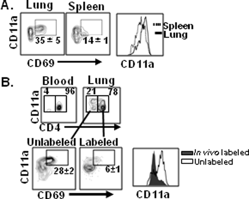
Figure 3. Distinct phenotype of tissue-resident versus circulating memory CD4 T cells.

(A) Cell surface CD11a and CD69 expression by lung- and spleen-derived HA-specific memory CD4 T cells gated on live CD4+CD44hiCD62Llo T cells, and are representative of 4 independent experiments. (B) In vivo labeling delineates resident and circulating polyclonal lung memory CD4 T cell subsets. BALB/c mice previously infected with influenza (4–6 weeks post-infection) were injected intravenously with fluorescently labeled anti-CD4 antibody, and blood and lung tissue were harvested. Upper: Proportion of CD3ε+CD8α− γδ− T cells stained by or protected from in vivo administered antibody. Lower: CD11a and CD69 expression by the labeled or protected cell populations, representative of 4 independent experiments.
