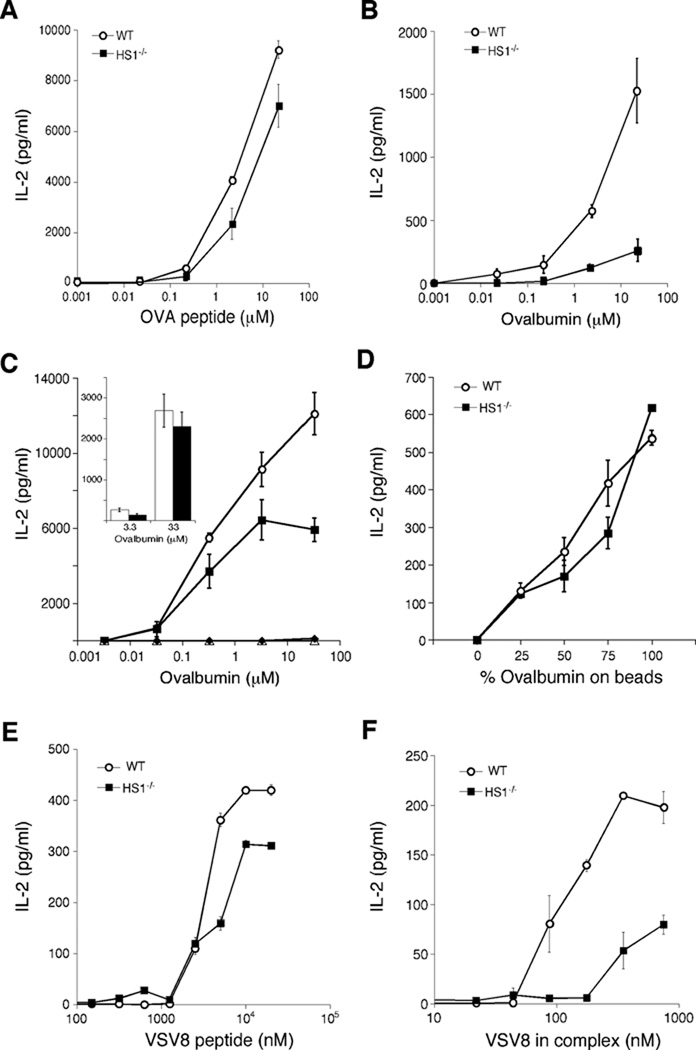
Fig. 2. HS1 is required for presentation of protein antigens.
(A–B) BMDCs cultured from WT and HS1−/− on the Balb/c background were pulsed with the class II-restricted OVA323–339 peptide (A) or with the whole ovalbumin protein (B) at the indicated doses for 4h. They were then co-cultured with OVA-specific DO-11.10 T cells and 24h later, IL-2 levels in the culture supernatants were measured by ELISA. (C) BMDCs cultured from WT and HS1−/− mice on the C57Bl/6 background were incubated in the presence or absence of mannan to block mannose receptors, and then ovalbumin protein was added, so that any residual uptake would be by macropinocytosis. Antigen presentation to OTII T cells was then assayed as in panels A and B. In this experiment (representing 2 of 4 replicates), mannan completely blocked the response, indicating that even very high doses of ovalbumin are taken up almost exclusively by receptor-mediated endocytosis.  , WT DCs without mannan; ■, HS1−/− DCs without mannan; Δ, WT DCs with mannan; ◆, HS1−/− DCs with mannan. Inset, data from a separate experiment (representing 2 of 4 replicates) where T cell responses to mannan-blocked DCs were measurable at high ovalbumin doses. Open bars, WT DCs with mannan, filled bars, HS1−/− DCs with mannan; differences between WT and HS1−/− DCs in the presence of mannan were not statistically significant at any dose of ovalbumin. (D) WT and HS1−/− BMDCs (C57Bl/6 background) were allowed to phagocytose latex beads coated with ovalbumin and serum albumin in the indicated ratios, holding total protein and bead number constant. OTII T cell responses were then measured. (E–F) WT and HS1−/− BMDCs were pulsed with free VSV8 peptide (E) or VSV8 peptide pre-bound to GRP94 (F) at the indicated doses for 4h. VSV8-specific N15 T hybridoma cells were incubated with antigen-pulsed BMDCs for 24h and IL-2 levels in the supernatants were measured by ELISA. Data represent means +/− StDev from replicate wells of one representative experiment.

