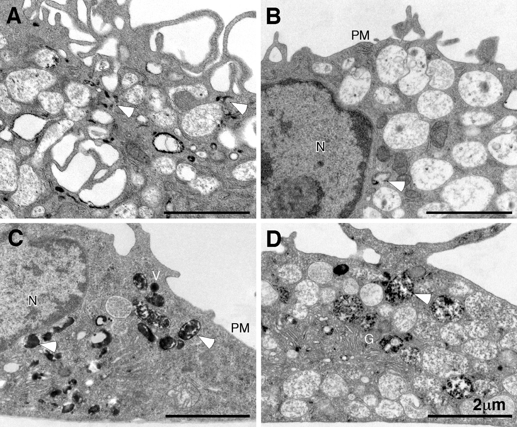
Fig. 5. Analysis of fluid-phase and receptor-mediated endocytosis by electron microscopy.
WT (A, C) or HS1−/− (B, D) BMDCs were incubated with transferrin-HRP (A, B) for 15 min at 37°C to load endocytic compartments via receptor-mediated endocytosis. Alternatively, BMDCs were incubated with soluble HRP (C, D) for 30 min at 37°C, to load the endocytic pathway via fluid phase endocytosis (macropinocytosis). Cells were fixed and processed with DAB to generate an electron-dense reaction product and visualized by transmission electron microscopy. Representative images from 3 independent experiments are shown. Arrowheads indicate compartments (early endosomes and macropinosomes) filled with electron-dense HRP reaction product. Note the relative paucity of electron-dense deposits in the HS1−/− cells when HRP is delivered by receptor-mediated endocytosis (compare A and B), but not macropinocytosis (compare C and D). N, nucleus; PM, plasma membrane; G, Golgi complex; V, macropinocytic vacuoles.

