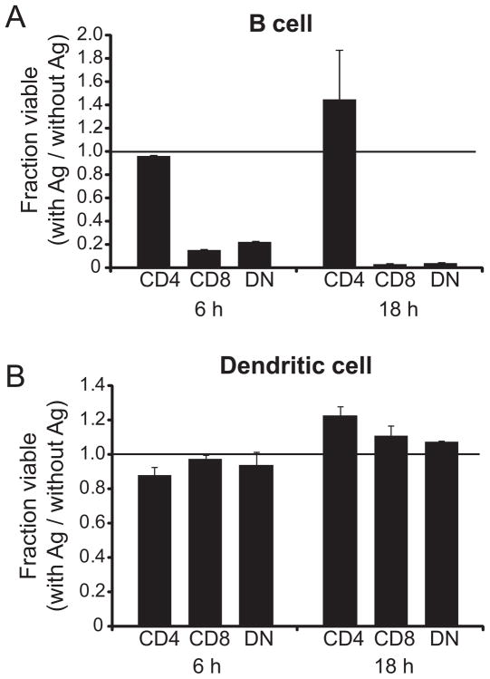
Figure 5. Ag specific cytolysis by G107S CD8+ and DN T cells.
Purified CD4+, CD8+ and DN G107S T cells were stimulated for 4 days with MOG35–55 Ag, isolated, and then co-cultured with purified B6 B lymphocytes (A) or BM-derived DCs (B) at a 1:1 E:T ratio in triplicate. Cultures were either pulsed with 100 μg/ml MOG35–55 or left without Ag. At 6 or 18 h numbers of residual viable target cells was quantitatively assessed by flow cytometry. The ratio of residual cells in Ag-pulsed/non-pulsed cultures is plotted. Mean + 1 s.d. is shown. Representative of 2 experiments.

