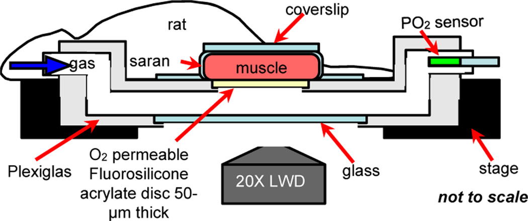
Figure 1.
IVVM classical experimental setup. Extensor Digitorum Longus (EDL) muscle of the rat is surgically exposed and is positioned surface down on the viewing platform of an inverted light microscope. Oxygen is delivered to the muscle using a gas exchange chamber connected to computer-controlled flow meters. Oxygen levels in the chamber are measured and monitored using a fibre optic oxygen sensor probe. The tissue is trans-illuminated and blood flow responses are recorded and analyzed as oxygen levels are simultaneously oscillated.

