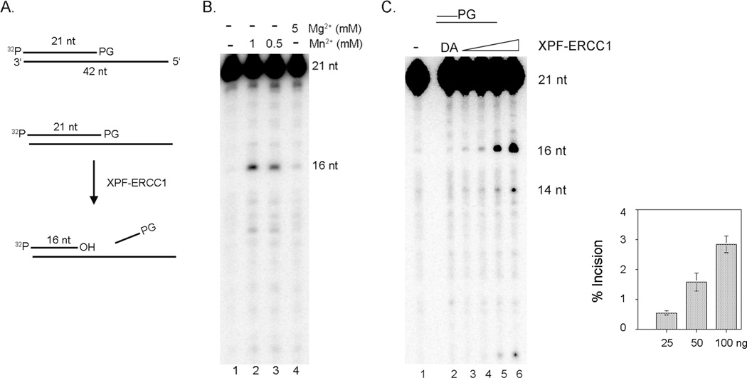
Figure 2. XPF-ERCC1 removes a 3’-phosphoglycolate (3’-PG) at a 3’-terminus in vitro.
(A). Substrate DNA and in vitro nuclease assay A 21 nt oligonucleotide with a PG at the 3’ end was labeled with 32P and annealed to a partially complementary 42 nt oligonucleotide to generate the DS/SS substrate. Incision products by XPF-ERCC1 are analyzed on a 10% sequencing gel. (B). Mn2+ stimulates the removal of 3’-PG by XPF-ERCC1. The 5’-labeled DS/SS substrate with a 3’-PG (2 nM) was incubated with XPF-ERCC1 (35 nM) at 30°C for 1 hr. XPF-ERCC1 released 5 nt fragments from the 3’end by removing 3’-PGs. Lane 1, no XPF-ERCC1; lane 2, with 1 mM MnCl2; lane 3, with 0.5 mM MnCl2; and lane 4, with 5 mM MgCl2. 2.8 %, 2.2 % and 0.8 % of the substrate was incised in lanes 2, 3, and 4, respectively. (C). Removal of 3’-PG by XPF-ERCC1. Increasing amounts of XPF-ERCC1 were incubated with 5’-labeled DS/SS substrate (2 nM) with a 3’-PG in 10 µl reaction buffer at 30°C for 1 hr. Lane 1, no XPF-ERCC1; lane 2, the endonuclease defective XPF(DA)-ERCC1 (42 nM); lane 3, 7 nM; lane 4, 14 nM; lane 5, 28 nM; lane 6, 56 nM. No incision was detected in lane 2. 0.4%, 0.6%, 1.5%, and 3% were incised in lanes 3, 4, 5, and 6, respectively. The average incision activities at the indicated amount of XPF-ERCC1 from three different experiments were plotted in a graph with standard deviations (error bars).

