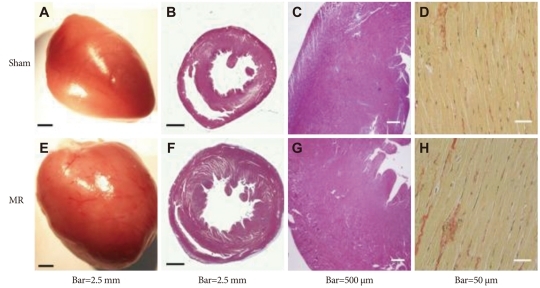
Fig. 6.
Comparison of pathological results. Eccentric hypertrophy developed (E) and left ventricular mass increased (F). Masson's trichrome staining showed no significant difference in interstitial fibrosis (B, F, C and G). Picrosirius red staining showed similar collagen content in both groups (D and H).

