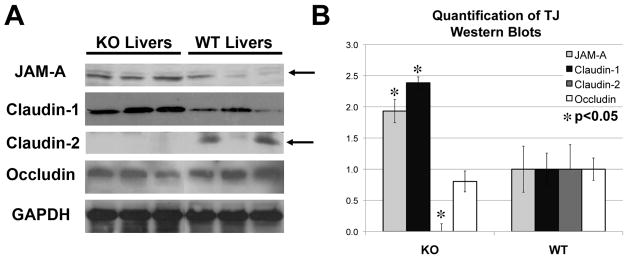
Figure 5. Changes in TJ protein expression in the KO livers.
A. Examination of TJ protein expression by western blots using whole-cell lysates shows an increase in JAM-A (35kDa) and claudin-1 (22kDa), no change in occludin (60–82kDa) and absence of claudin-2 (22kDa) in KO livers. GAPDH served as the loading control.
B. Normalized (to GAPDH) densitometric analysis reveals significantly (P <0.05) lower average (+/−SD) protein expression of claudin-2 and higher average (+/−SD) expression of JAM-A and claudin-1 in KO than WT livers (n≥3), while occludin remained unchanged.

