
Fig. 1.
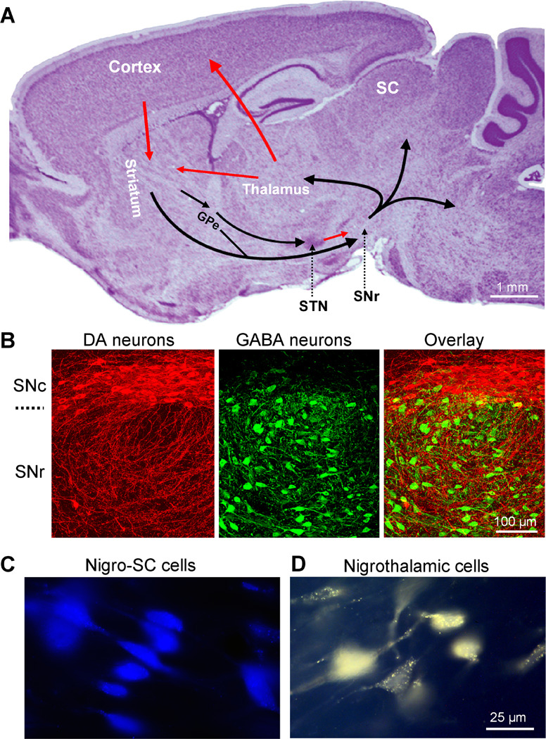
The substantia nigra pars reticulata (SNr) is a key output nucleus of the basal ganglia. (A) A Nissl-stained sagittal mouse brain section showing the location of SNr and other major components of the basal ganglia. Arrows indicate information flow directions. The SNr receives information from several components of the basal ganglia and sends output to the thalamus, superior colliculus (SC), and brainstem motor structures. Unpublished data of FMZ. (B) Confocal images of double immunohistochemical staining for tyrosine hydroxylase (TH, red, a key enzyme for dopamine synthesis) and parvalbumin (PV, green, expressed only in GABA neurons). Most TH-positive dopamine neurons are in SNc with their dendrites extending into SNr where most neurons are PV-positive GABA neurons. Modified from Zhou et al. 2009 with permission. (C) Examples of retrogradely labeled SNr neurons projecting to the superior colliculus (SC). Blue is a pseudocolor in this image for easier visualization. (D) Examples of retrogradely labeled SNr neurons projecting to the thalamus. C and D are modified from Lee and Tepper, 2007a with permission. Copyright 2006 Wiley-Liss, Inc.