
Fig. 3.
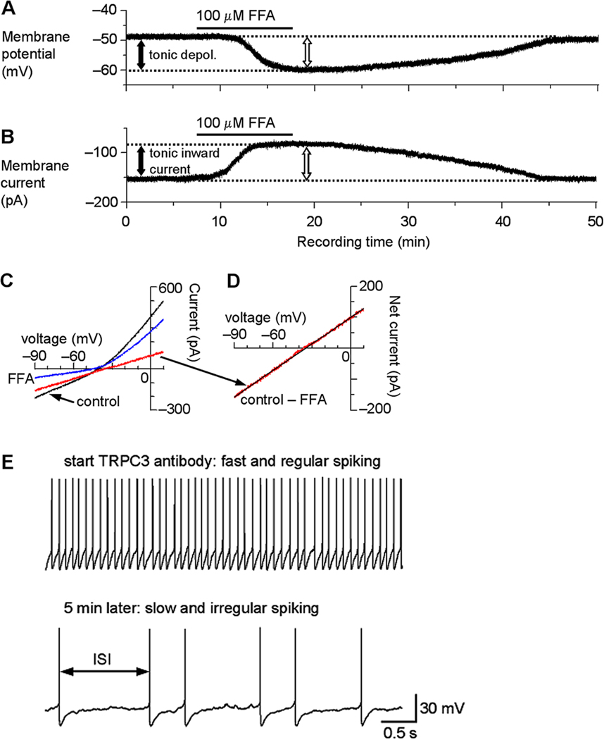
TRP channels mediate a tonic inward current and depolarization in SNr GABA neurons. (A) After blocking NaV channels with 1 µM TTX and under current clamp recording condition, bath application of 100 µM flufenamic acid (FFA) induced a hyperpolarization of about 10 mV (open arrow), indicating an FFA-sensitive tonic depolarization (depol., filled arrow). (B) When voltage clamped at −70 mV, 100 µM FFA induced an outward current (open arrow), or reduced a potential tonic inward current, as reflected by the apparent reduction of the holding current (filled arrow). (C) A linear voltage ramp from −90 mV to 10 mV was applied under control condition (black trace) and during 100 µM FFA application (blue trace). Digital subtraction (control – FFA) revealed the current inhibited by FFA (red trace) and its I–V relationship. The decreased current also indicates an increased input resistance or decreased whole cell conductance. (D) The FFA-inhibited current in C displayed at an enhanced scale. The I–V relationship is clearly linear with no signs of voltage-dependent activation or inactivation and reversed its polarity around −35 mV on average (intercept on X-axis in linear regression analysis as indicated by the black straight line). (E) Intracellular application of a TRPC3 antibody decreases the firing rate and increases the firing irregularity in SNr GABA neurons. Within the first minute of recording when the antibody was not likely to have diffused sufficiently into the cell, the firing was fast around 10 Hz and had a regular inter-spike interval (ISI). During the fifth minute of recording when a considerable amount of the antibody was likely to have diffused into the cell, the spontaneous firing became much slower and had an irregular ISI. Modified from Zhou et al., 2008 with permission.