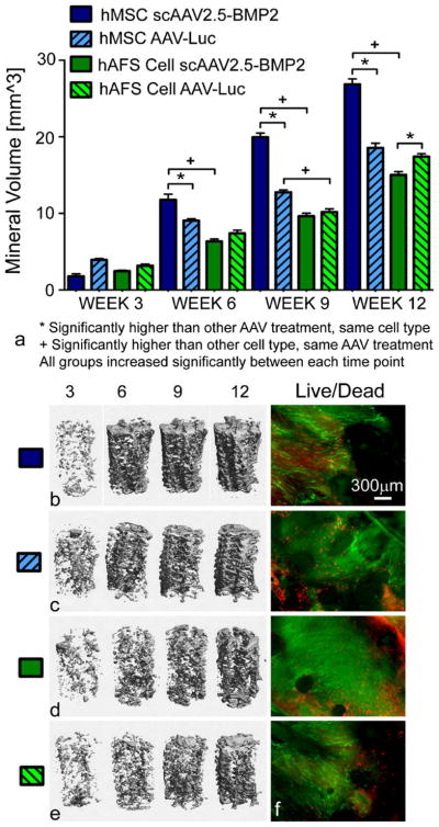
Fig. 5.
Evaluation of osteogenic differentiation of stem cells seeded on three-dimensional PCL scaffolds previously coated with lyophilized AAV. a Quantitative comparison of mineral volumes within PCL scaffolds. *,+P<0.05. b–e Representative micro-CT images of mineral formation within scaffolds. f Live/Dead microscopy images of scaffolds showing live green cells along the circumferential periphery of scaffolds (red dead cells). Images are shown at ×4 magnification

