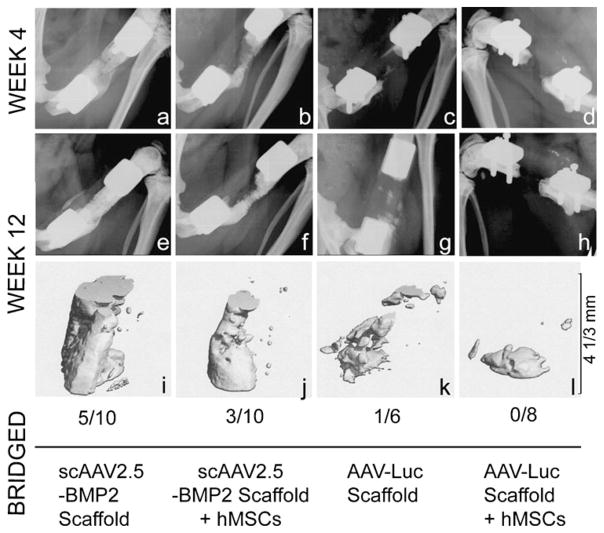
Fig. 7.
Qualitative mineral formation at the defect site after in vivo delivery of AAV-coated PCL scaffolds with or without pre-seeding of hMSCs. Radiographic (a–h) and in vivo micro-CT (i–l) images from defects that had the representative mineral formation per group, together with the bony bridging rate for each treatment group

