


Figure 2.
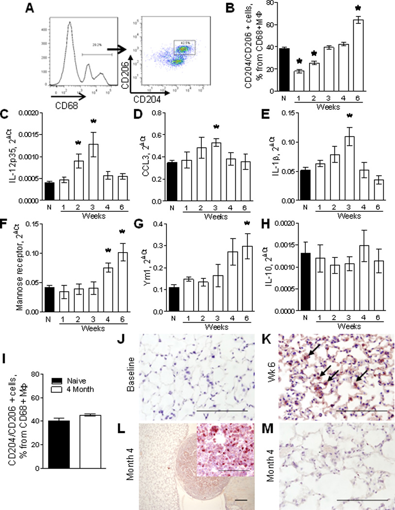
Characterization of macrophage phenotype in lungs following urethane treatment. A) Representative FACS plots demonstrating identification of M2 polarized macrophages in whole lungs of a naïve (untreated) mouse using anti-CD204 (scavenger receptor) and anti-CD206 (mannose receptor) antibodies. B) Analysis of the percentage of CD204 and CD206 double-positive cells within CD45+CD68+ macrophages in lungs of naïve mice (N) and mice at week 1–6 after urethane treatment. C–H) mRNA expression for M1 markers IL-12p35, CCL3, IL-1β and M2 markers mannose receptor, Ym1, IL-10 in total CD11b-positive cells isolated from lungs of naïve mice and 1–6 weeks after a single injection of urethane. n=4 mice per time point, *= p<0.05 compared to naïve mice. I) Percentage of CD204 and CD206 double-positive cells within lung CD45+CD68+ macrophages at 4 months after urethane injection. n=4 mice per group. J–M) Representative photomicrographs of lung sections stained with anti-arginase-1 antibodies at different time points after injection of urethane. Bars: 100µM.