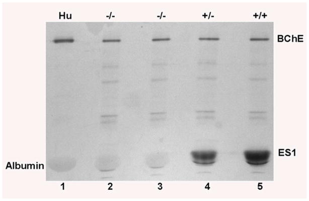
Figure 3.
Visualization of ES1 activity in the plasma of ES1+/+, +/− and −/− mice using α-NA as the substrate. Plasma was loaded at 2 μl/well onto a nondenaturing gradient polyacrylamide gel. Lane 1) Human plasma has BChE and albumin pseudo-esterase activity, but no carboxylesterase. Lanes 2 and 3) Plasma from ES1−/− mice demonstrates the complete absence of carboxylesterase activity in these mice. Lane 4) ES1+/− plasma. Lane 5) ES1+/+ plasma. The ES1 band intensity is greater in lane 5 than lane 4, corresponding to the higher carboxylesterase activity in +/+ than +/− plasma.

