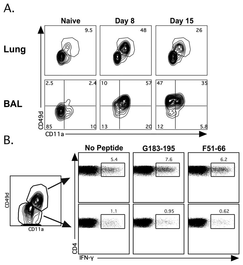
Figure 5.
CD11a and CD49d identify Ag-specific CD4 T cells following acute RSV infection. BALB/c mice were infected with RSV i.n.. (A) Representative plots depict CD11a and CD49d expression on lung and BAL CD4+Thy1.2+-gated CD4 T cells at day 8 and 15 post-infection. (B) Lung CD4 T cells from day 8 RSV infected BALB/c mice were incubated in BFA alone or stimulated with either the RSV peptide G183–196 or F51–66. Representative plots depict IFN-γ production between CD11aloCD49d− (top) and CD11ahiCD49d+ (bottom) CD4 T cells (CD4+CD90.2+). Similar results were obtained from 4 independent experiments with 3–4 mice per group.

