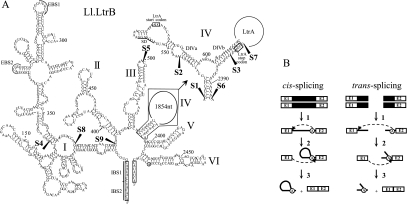
FIGURE 1.
Ll.LtrB secondary structure and splicing pathways. (A) Ll.LtrB secondary structure. The six domains of Ll.LtrB are indicated (I–VI) and the detailed secondary structure of a portion of domain IV is also shown (top right). The ltrA start and stop codons are boxed and the Shine–Dalgarno sequence (SD) is underlined. Exons 1 and 2 at both 5′ and 3′ extremities of the intron are also boxed. Fragmentation sites are mapped with black arrowheads (S1–S9). S4 was not used in this study. EBS1 and EBS2, exon-binding site 1 and 2; IBS1 and IBS2, intron-binding site 1 and 2; branch point, circled A. (B) Group II intron cis- and trans-splicing pathways. Following transcription of the interrupted gene (step 1), the 2′-OH of the bulged adenosine present in domain VI (circled A) performs the initial nucleophilic attack at the exon 1–intron splice junction generating a 2′–5′ linkage (step 2). Then, the 3′-OH of the released exon 1 performs the second nucleophilic attack at the intron–exon 2 splice junction, releasing the intron lariat and ligating the flanking exons (step 3). Fragmented group II introns can also splice in trans using the same pathway (steps 1–3). Because trans-splicing occurs between two independent RNA transcripts, the intron is released as a Y-branched molecule instead of a lariat. Group II intron, black line; exon 1 and 2, E1 and E2; branch point, circled A.

