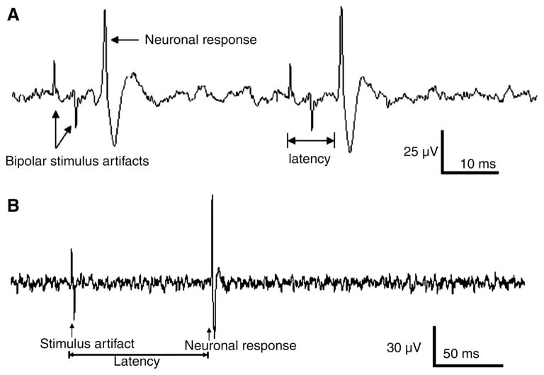
Fig. 4.
Vagal nodose ganglia neuronal responses to electrical stimulation of subdiaphragmatic vagal afferent nerves (SDVAS). A: action potential elicited from a nodose ganglia neuron in response to 2 consecutive electrical subdiaphragmatic vagal stimulations (100 μA, 20 Hz). Conduction distance = 0.06 m, latency = 0.007 s, conductive velocity = 8.5 m/s, suggesting that we were recording a gastrointestinal Aδ fiber. B: action potential elicited from a nodose ganglia neuron in response to high-intensity electrical vagal stimulation (EVS) (400 μA, 1 Hz). The conductive distance = 0.06 m, latency = 0.07 s, conduction velocity = 0.8 m/s, suggesting that a gastrointestinal C fiber was recorded.

