
Figure 3. Sequence comparison of RIG-I orthologs and related RLR proteins.
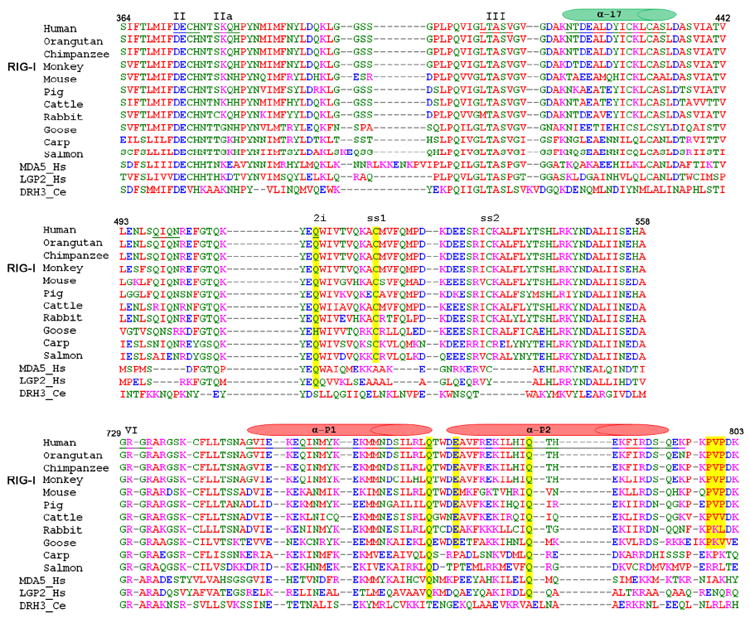
Selected sequence alignments of all available RIG-I orthologs (top 11 rows) and comparison with closely related RLR proteins MDA-5 and LGP-2 from Homo sapiens, Dicer-related-Helicase 3 (DRH-3) from Caenorhabditis elegans. Top panel shows conservation of Motif IIa and the a17 helical shaft (green cylinder). Middle panel shows conservation of insertion domain HEL2i. Bottom panel shows conservation of residues in Pincer domain, and helices of the pincer are indicated (red cylinders). Yellow lines indicate positions of mutated residues. A complete alignment of RLR sequences is provided as Fig. S2.