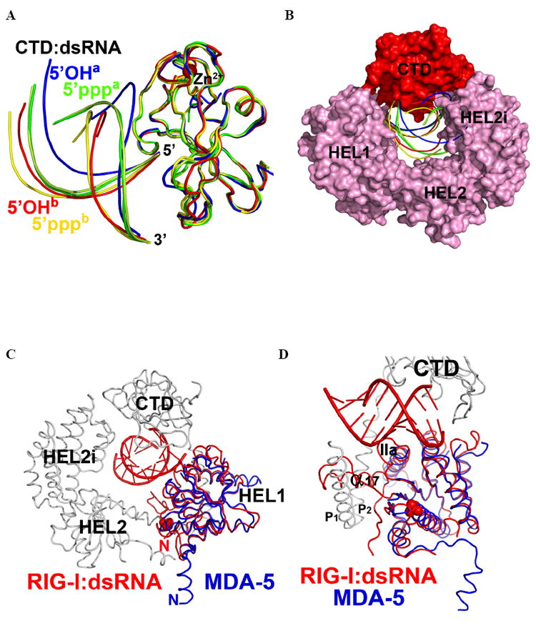
Figure 4. Superposition of RLR CTD-dsRNA complexes.

Comparison of the RNA duplex orientation in RIG-I (ΔCARDs):dsRNA with the available CTD:dsRNA structures. (A). CTDs of all complexes are superimposed and color-coded as follows. Red: CTD of RIG-I (ΔCARDs) and its complex with 5’OH-dsRNA10 complex, designated “5’OHb”. Green: RIG-I CTD and 5’ppp-dsRNA12 (PDB: 3LRR), designated 5’pppa. Pale Green: RIG-I CTD and 5’ppp-dsRNA12 (PDB: 3NCU), labeled as “5’pppa”. Blue: RIG-I CTD and 5’OH-dsRNA14 (PDB: 3OG8), labeled as “5’OHa”. Yellow: RIG-I CTD and 5’ppp-dsRNA14 (PDB: 3LRN), labeled as “5’pppb”. (B). Space-filling representation of RIG-I (ΔCARDs):dsRNA showing variation in RNA position among superimposed structures (C) Superposition of the RIG-I (ΔCARDs) and 5’OH-dsRNA10 complex and the MDA-5 HEL1 (blue ribbon) (PDB: 3B6E). (E) Close-up view of (D). See also Fig. S4.
