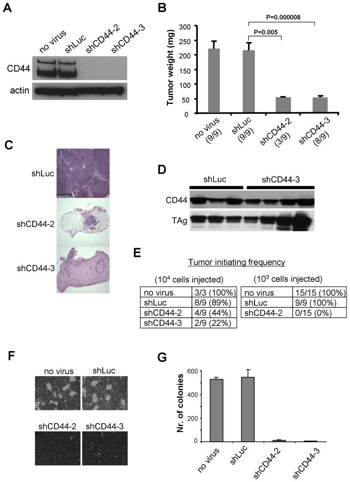
Figure 5. Tumor Growth of Human Mammary Epithelial BPLER Cells with Normal and Suppressed CD44 Expression.
BPLER cells were infected with a lentiviral vector expressing an shRNA to firefly luciferase (shLuc) or to CD44 (shCD44-2 and shCD44-3). The infection efficiency was more than 95% for all constructs (data not shown).
(A) The level of CD44 expression in individual infected cell populations in vitro was analyzed by western blotting 72h after the infection.
(B) Individual lentivirus-infected cells (106 cells per injection) were injected in nude mice and tumor weights (mean±SEM) were analyzed 4 weeks after the injection. Tumor incidence per injection is indicated in parentheses.
(C) Hematoxylin and eosin staining of the resulting tumors. Scale = 500μm.
(D) The level of CD44 expression in tumors arising from individual infected cell populations was analyzed by western blotting 72h after the infection.
(E) Tumor-initiating frequency of individual infected cell populations.
(F, G) Soft agar colony assay of individual lentivirus-infected cells. The cells (105/6cm dish) were plated in culture medium in soft agar and cultured for 4 weeks. The assay was terminated when colonies of control cells reached 1mm in diameter, at which point they were counted (mean±SEM, n+3).

