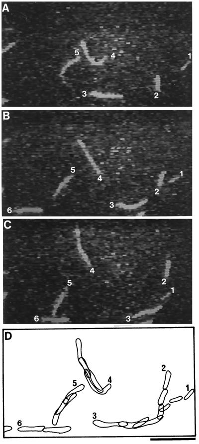
Figure 1.
Sequential photographs (A–C) of moving RP-labeled F-actins over a glass surface coated with 170-kD myosin in the presence of EGTA. Photographs (A–C) were taken at time intervals of 0.66 s. Traces of the six RP-labeled F-actins (1–6) shown in A to C are superimposed in D. The bar represents 10 μm.

