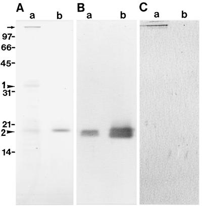
Figure 5.
Immunoblotting of the 170-kD myosin fraction (a) and spinach CaM (b). The concentrations of 170-kD myosin and CaM applied on SDS-PAGE for each assay were 0.9 and 0.2 μg, respectively. A, Coomassie brilliant blue staining of a 15% polyacrylamide gel. B, Immunoblotting using antiserum against spinach CaM. C, Immunoblotting using antiserum against the 170-kD heavy chain. The arrow indicates the position of the 170-kD heavy chain. Arrowheads 1 and 2 indicate the 34- and the 18-kD polypeptide, respectively. The Mrs (×10−3) of standard proteins are indicated on the left.

