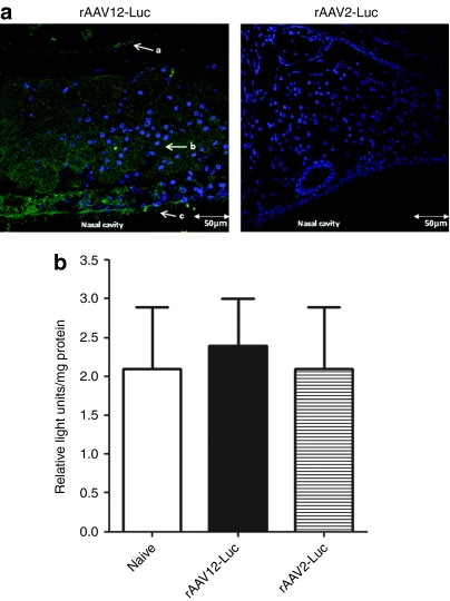
Figure 3.
Detection of luciferase expression by immunofluorescence in the anterior nasal cavity tissue of recombinant adeno-associated virus type 12 (rAAV12)-Luc treated mice. Mice were administered either 1 × 10¹¹ rAAV12-Luc or rAAV2-Luc vector particles by the intranasal (IN) route. Five days after rAAV-Luc administration, two mice from each group were sacrificed, and nasal cavities were excised. (a) Cross-sections of nasal cavity tissue were immunostained for the presence of luciferase as described in the methods. Nuclei were stained with 4′,6-diamidino-2-phenylindole (DAPI). Sections were viewed by confocal microscopy and images captured at ×40 objective. Arrow a indicates respiratory epithelium surrounding nasoturbinate bone, arrow b indicates lamina propria with Bowman's glands, and arrow c indicates ciliated pseudostratified olfactory epithelium. Images are representative of luciferase staining in both mice sacrificed from each group. Bars = 50 µm. Cross-sections of nasal cavity tissue of naive mice were stained for luciferase expression as a negative control (data not shown). (b) To confirm lack of expression in the lungs of rAAV-Luc treated mice, ex vivo luciferase assay was performed on homogenized lung tissue and expression was measured as described in the Materials and Methods section. Tissue from a naive mouse was included as a negative control. Error bars represent the mean relative light units (RLU)/mg protein + s.e.m., where n = 3. Statistical comparisons were performed by unpaired t-tests.

