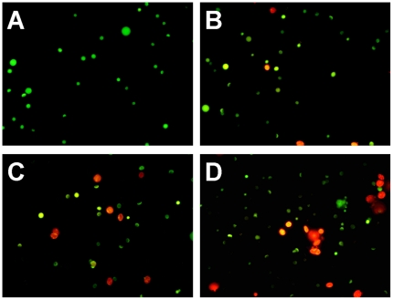
Figure 3. Effects of Cd on the testis of Sinopotamon henanense by fluorescence microscope.
(A) Control group, (B) Treated with 7.25 mg·L−1 Cd group, (C) Treated with 29.00 mg·L−1 Cd, (D) Treated with 116.00 mg·L−1 Cd. Three types of cells can be recognized: live cells (green), live apoptotic cells (yellow), and dead cells by necrosis (red). Compared with the control group, the number of apoptotic cells were significantly increased with increasing Cd concentrations.

