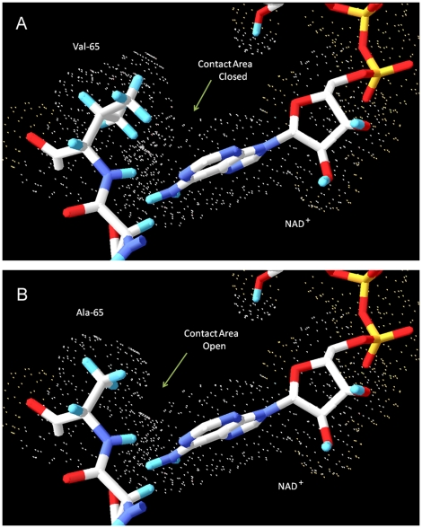
Figure 4. Van der Waals interactions between the adenine ring of NAD+ and the side chain of residue 65 of HSD10.
The wild type HSD10 and mutant HSD10 were shown in part (A) and (B), respectively. Different colors represent different atoms: carbon (white), hydrogen (blue), nitrogen (purple), phosphorous (yellow) and oxygen (red). For clarity, all other amino acid residues in the protein, other than the neighboring aspartate 64 have been rendered invisible. Small dots represent the extent of the van der Waals radii for atoms in amino acid residue 65 in the protein and in the NAD+.

