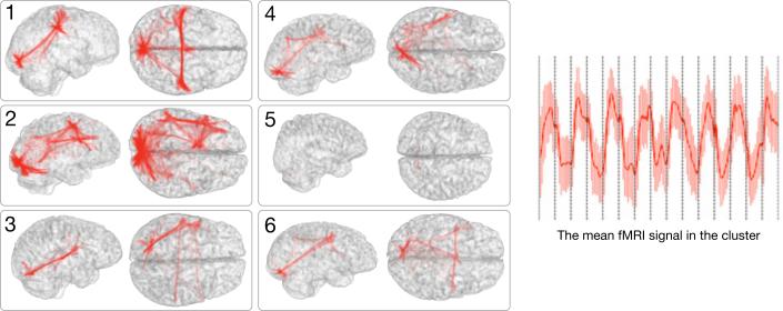
Fig. 3.
A cluster in the joint map corresponds to a network in each subject. Here we illustrate a network in 6 subjects that corresponds to one cluster in the atlas, and the mean fMRI signal of this cluster. The 8 block stimulus in this language study was not used by the analysis, but was recovered by the algorithm and corresponding networks were identified across all subjects. They typically span the visual cortex, the language areas (Wernicke and Broca), and the motor areas in some cases.

