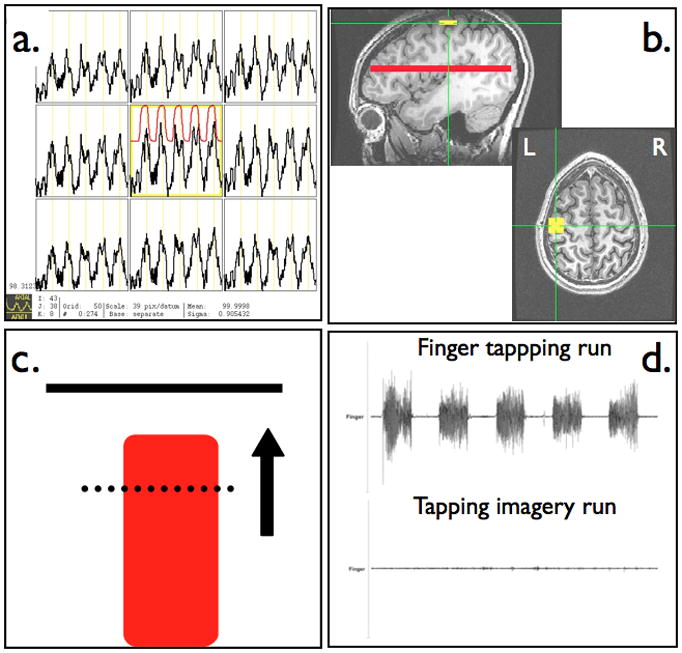
Figure 1.

Real-time fMRI scanning protocol with (a) continuously updated BOLD time series used for functional localization of primary motor cortex, (b) placement of square primary motor cortex ROI and whole brain axial slice for a reference ROI, (c) continuously updated neurofeedback display reflecting scan-to-scan level of BOLD signal within motor cortex ROI, and (d) monitoring of muscle movement during finger tapping and motor imagery neurofeedback tasks.
