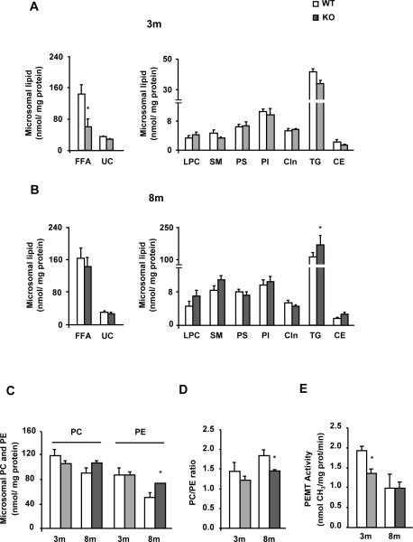
Fig. 3. MAT1A deletion alters the microsomal lipid composition in mice.
Liver microsomes were isolated from 3-month-old (3m) and 8-month-old (8m) wild type (WT) (□) and MAT1A-knockout (MAT1A-KO) ( ) mice by differential centrifugation. (A–D) Lipids were extracted from liver microsomes, and free fatty acid (FFA), unesterified cholesterol (UC), lysophosphatidylcholine (LPC), sphingomyelin (SM), phosphatidylserine (PS), phosphatidylinositol (PI), cardiolipin (Cln), triglyceride (TG), cholesteryl ester (CE), phosphatidylcholine (PC) and phosphatidylethanolamine (PE) were separated and quantified as indicated in material and methods. (E) Microsomal phosphatidylethanolamine N-methyltransferase (PEMT) activity was determined by a radiometric assay. Values are means ± SEM of 4–8 animals per group. Statistical differences between MAT1A-KO and WT mice are denoted by *p<0.05 (Student's t test).

