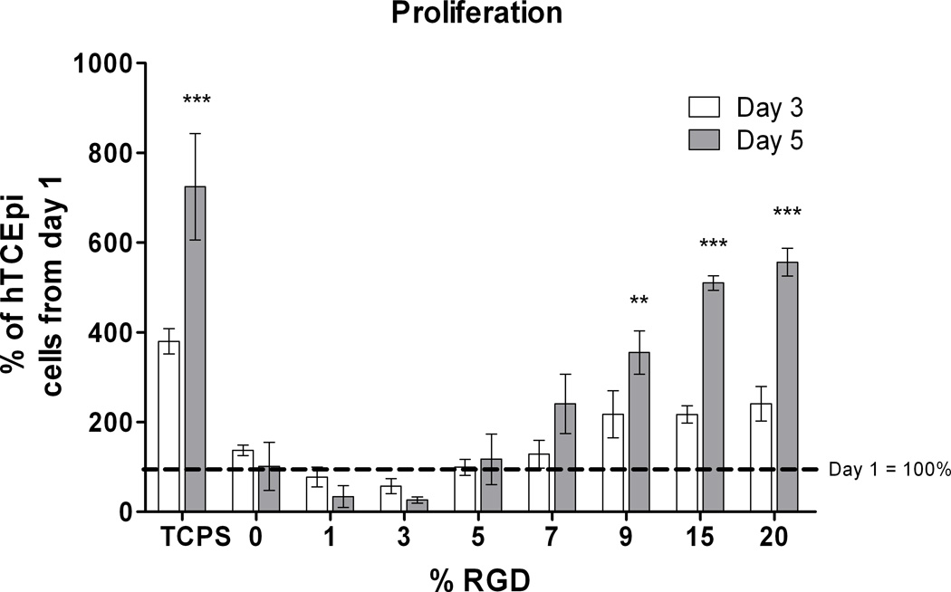
Figure 6.
hTCEpi cells demonstrated proliferation rates dependent on the surface density of RGD. On 0 to 3% RGD/d-glucamine films, cells did not attach well. Those cells that initially attached to the surface ultimately detached over the course of 5 days. Cells on surfaces fabricated with 5% RGD/d-glucamine did not proliferate, while cells on surfaces fabricated with 7% RGD/d-glucamine showed a slight increase in cell numbers. The highest rates of proliferation were observed on surfaces fabricated from 9–20% RGD/d-glucamine solutions. (*0.01 ≤ P < 0.05, **0.001 ≤ P < 0.01, ***P < 0.001 compared to 0, 1, 3, and 5 % RGD from day 5)

