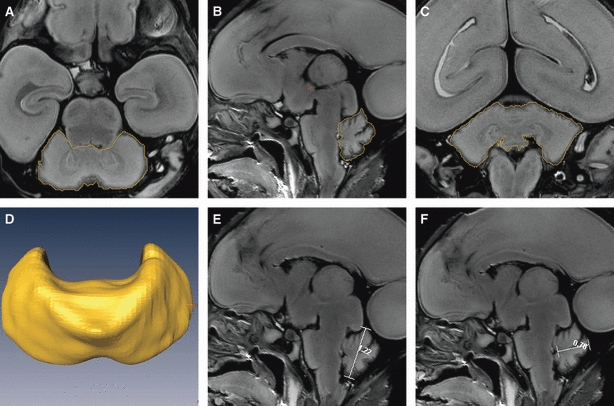
Fig. 1.

Procedures used to measure the CV, TCD, vermis height and vermis width by Amira. The figures show the procedures used to outline the cerebellum in a representative 21-week-old fetus in the (A) transverse, (B) sagittal and (C) coronal planes; the boundaries of the cerebellum are outlined in yellow. (D) The reconstructed cerebellar image after outlining. (E and F) The procedures used to measure the height and width of the vermis on median sagittal images by Amira; the lines on the images are the vermis height and width, respectively.
