
Fig. 7.
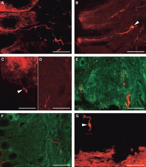
Transverse sections of pig gastric mucosa at the level of the diverticulum after the insertion of DiI crystals in the lamina propria. (A) Note a thin plexus of labelled fibres between the adenomers. (B) A neurone (arrowhead) in the lamina propria projecting to the epithelium. (C) Labelled neurones (arrowhead) projecting into a labelled follicle. (D,E) Labelled fibres in unlabelled follicles. (F) Labelled fibres resting on the connective follicular capsule. (G) Labelled, ovoid neurone (arrowhead) near the labelled muscularis mucosae. (E,F) Merging with the green filtre cube to best show the lymphoid follicle structure. Bars: (A,E,F) 200 μm; (B,C,G) 100 μm; (D) 50 μm.