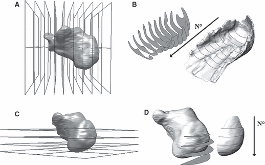
Fig. 3.

Generation of HFP sections. (A,B) Generation of coronal sections. (A) Identification of coronal planes. The step increment of 13% of calcaneus length was chosen to detect the plantar tuberosity of the calcaneus (i.e. the point where HFP thickness was measured in previous studies). (B) Extrapolation of HFP coronal sections. The coronal sections were numbered one to 10 starting from the most posterior section forward. (C,D) Generation of axial sections. (C) Identification of the axial planes. The step increment of 18% was chosen to detect the most posterior point of the calcaneus. (D) Extrapolation of HFP axial sections. To isolate the tissue behind the calcaneus, the posterior HFP was sectioned with the axial planes. The axial sections were numbered one to four starting from the most proximal section.
