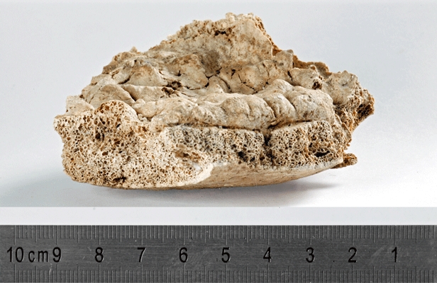
Fig. 8.

Hyperostosis frontalis interna: endocranial view of a frontal bone fragment before restoration. Note the clearly demarcated borders of the appositional bone adjacent to the sagittal sulcus and the continuity of the thickened diploe (left side of the pictures).
