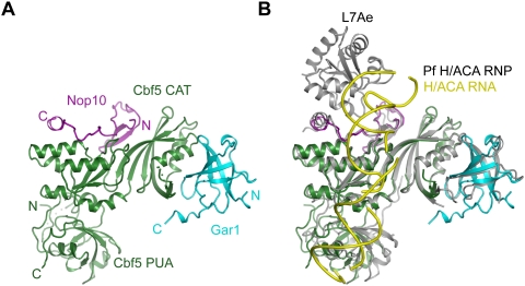
Figure 2.
Crystal structure of the yeast Cbf5C–Nop10–Gar1C complex. (A) Ribbon representation of the structure. Cbf5 is green, Nop10 is magenta, and Gar1 is cyan. The N and C termini of each protein are indicated. (CAT) The catalytic domain. (B) Structural alignment of the yeast Cbf5C–Nop10–Gar1C complex with the Pf H/ACA RNP. Pf H/ACA RNP structure is colored in gray for proteins and in yellow for RNA.

