Fig. 2.

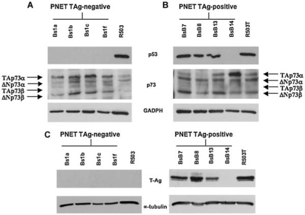
p53 and p73 expression in mouse T-antigen-negative (A) and -positive (B) mouse PNET cells and in mouse T-antigen-negative (R503; A) and -positive (R503/T; B) fibroblasts. The relative protein levels of p53 and p73 were detected by Western blotting using 40 μg of whole lysate. As shown in (A), the T-Ag-negative PNET cells do not express p53 but exhibit significant levels of TAp73α and ΔNp73α p73 isoforms. The T-Ag-positive PNET cells (B) express a prevalence of ΔNp73α and ΔNp73β isoforms. The presence of T-Ag in PNET cells was tested by Western blotting (C). The expression of GAPDH protein was assessed to normalize protein loading.
