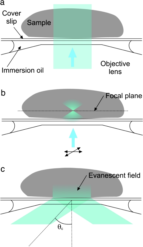
Fig. 1.
Comparisons between different fluorescence microscopy techniques. (a) Epifluorescence (EPI). The beam is incident along the central axis of the objective and illuminates the entire sample. Both in- and out-of-focus fluorescence signal is collected. (b) Laser confocal scanning microscopy (LSCM). The laser beam is focused in the plane of interest, reducing fluorescence excitation in other planes, and scanned across the sample. A pinhole (not shown) rejects most of the out-of-focus excited fluorescence and further confines the image to the focal plane of interest. (c) Total internal reflection fluorescence microscopy (TIRFM). A beam incident on the interface between two mediums (e.g. coverslip glass and water/sample) with different refractive indices at an angle greater than the critical angle θc is totally internally reflected. An evanescent field is generated, the intensity of which decays exponentially over a few hundred nanometres. (This figure is available in colour at JXB online.)

