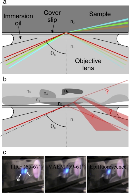
Fig. 4.
Distinguishing TIRFM and VAEM. (a) The angle at which the beam is incident on the refractive index interface determines whether the microscope operates in TIRFM or VAEM mode. The mode is easy to predict when only two refractive indices n1 and n2 are present. At angles greater than the critical angle θc (red line), the beam is totally internally reflected. If the angle is less than θc (green and blue lines), the beam is refracted and the microscope operates in VAEM mode. A tiny proportion of the beam, dependent on the angle, is reflected. If the beam is incident at the critical angle (orange line), then the width of the beam causes the microscope to operate in mixed modes. (b) It is difficult to predict the microscopy mode when multiple refractive indices are present within the complex geometry of a plant cell. (c) Visualizing the difference between TIRFM, VAEM, and epifluorescence modes using an Arabidopsis root. By temporarily placing a strip of sticky tape in the excitation light path it is possible to visualize the incident (arrow) and totally internally reflected beams simultaneously (arrow with star). In TIRFM the excitation beam returns strongly through the objective and is clearly visible on the sticky tape. In VAEM and standard epifluorescence microscopy, no more than a weak reflection returns through the objective.

