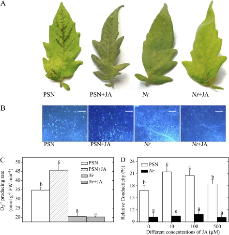
Fig. 3.
Effects of exogenous JA treatment on the sensitivity of detached PSN and Nr leaflets to AAL toxin. Detached leaflets were incubated under continuous light at 25°C for 48 h. (A) Symptoms of PSN and Nr leaflets after 0.2 μM AAL toxin application with or without 100 μM JA. (B) Callose deposition in PSN and Nr leaves in response to AAL toxin with or without 100 μM JA. Leaflets were stained with aniline blue to detect callose. Representative microscope images are shown. Callose deposits appear as bright spots on a dark background. Scale bars represent 100 μm. (C) Quantitative measurements of the O2·− level in PSN and Nr leaflets after AAL toxin application with or without 100 μM JA. (D) Quantitative measurements of relative conductivity in leaflets of PSN and Nr after AAL toxin application or co-treatment with the toxin and different concentrations of JA. Error bars indicate standard deviation. For each figure, letters indicate significant differences among treatments (P <0.05, Duncan's multiple range test). (This figure is available in colour at JXB online.)

