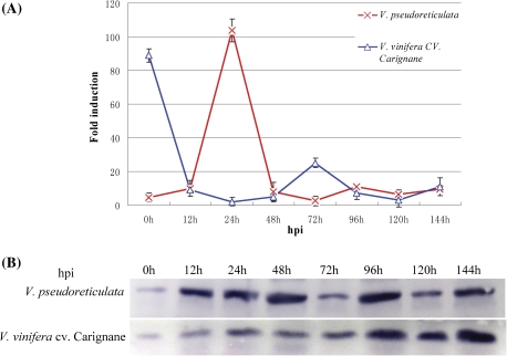
Fig. 2.
Expression of RFP1 in grapevine leaves that were inoculated with the pathogen U. necator. Leaves of plantlets of V. pseudoreticulata accession Baihe-35-1 and V. vinifera cv. Carignane were inoculated with U. necator (5×10⁵ spores ml−1). Control leaves were sprayed with sterile water. Leaves were collected at different time points as indicated. Hpi, hours post-inoculation with fungal spores. (A) Analysis of VpRFP1 expression at the transcript level by real-time PCR. VpGAPDH was used as the internal control. Data are the mean ±SE (n=3) of a representative experiment. (B) Analysis of VpRFP1 expression at the translational level by western blot. A 28 μg aliquot of total protein was used for each line, separated by SDS–PAGE, transferred onto a PVDF membrane, and immunodetected with a VpRFP1-specific antiserum. (This figure is available in colour at JXB online.)

