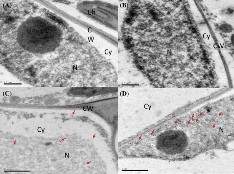
Fig. 3.
Immunogold localization of VpRFP1 in Vitis pseudoreticulata leaves. (A) With use of the rabbit pre-immune serum instead of the specific rabbit antiserum as control, no substantial signal was detected. (B) The absence of the antiserum to test the non-specific labelling of goat anti-rabbit IgG antibody–gold conjugate; no substantial signal was detected. (C) Before inoculation with pathogen, a few gold particles were detected in the nucleus and cytoplasm. (D) At 24 h after inoculation with pathogen, more gold particles were detected in the nucleus. N, nucleus; Nu, nucleolus; Cy, cytoplasm; CW, cell wall. (This figure is available in colour at JXB online.)

