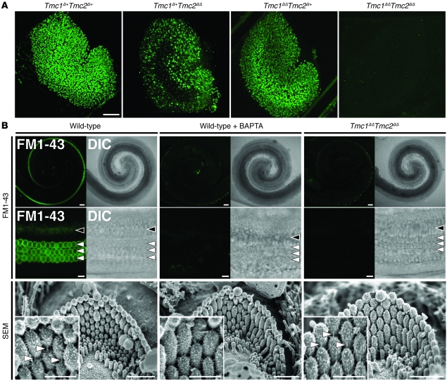
Figure 8. Tmc1Δ and Tmc2Δ hair cell uptake of FM1-43.
(A) Confocal microscopic images of P3 mouse utricles (Tmc1Δ/+Tmc2Δ/+, n = 4 utricles; Tmc1Δ/+Tmc2Δ/Δ, n = 6; Tmc1Δ/ΔTmc2Δ/+, n = 4; Tmc1Δ/ΔTmc2Δ/Δ, n = 6) exposed to 5 μM FM1-43. Scale bar: 100 μm. (B) IHCs and OHCs of P3 wild-type mice (n = 5 mice) had robust uptake of FM1-43. Wild-type hair cells (n = 5 mice) pretreated with 5 mM BAPTA did not take up FM1-43, and tip links are missing (tip links indicated by white arrowheads in insets). No FM1-43 uptake was detected in Tmc1Δ/ΔTmc2Δ/Δ hair cells (n = 5 mice) despite intact hair bundles and tip links (compare insets). Black arrowheads, IHCs (middle turn); white arrowheads, OHCs (middle turn); DIC, differential interference contrast. Gamma settings for green channel were adjusted equally throughout entire images and for all images using Adobe Photoshop CS5 software. Scale bars: 100 μm, upper panels; 10 μm, middle panels; 1 μm, lower panels; 500 nm, insets in bottom row of panels. See also Supplemental Figure 6.

